Radiology
Radiology, also known as diagnostic imaging, is a series of tests that take pictures of images of parts of the body. The field encompasses two areas - diagnostic radiology and interventional radiology.
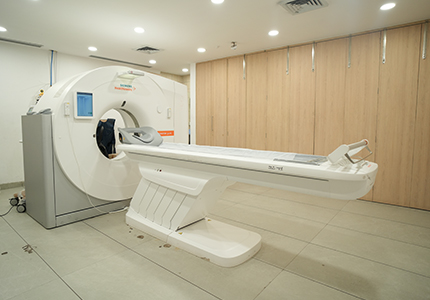
A variety of imaging techniques such as Radiography, Ultrasonography, Computed Tomography (CT), Nuclear Medicine including Positron Emission Tomography (PET), Fluoroscopy, and Magnetic Resonance Imaging (MRI) are used to diagnose or treat diseases. Interventional radiology is the performance of usually minimally invasive medical procedures.
CBCT
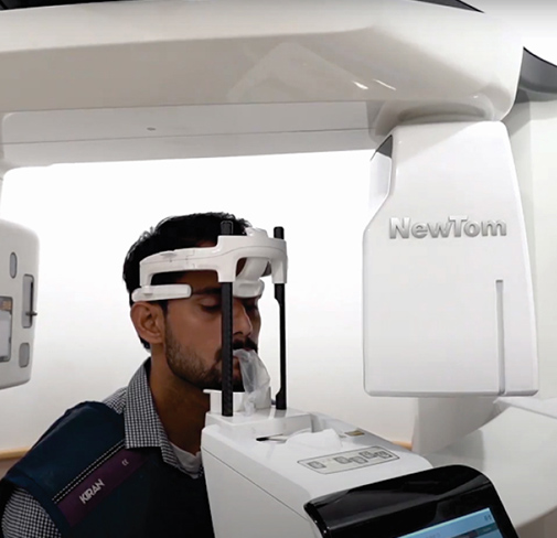
CBCT (Cone Beam Computed Tomography) scan is a specialized imaging technique that generates detailed three-dimensional images of various structures in the body, particularly the head, face, and dental structures. It is a type of computed tomography (CT) scan that uses a cone-shaped X-ray beam to capture multiple images from different angles, which are then reconstructed into a 3D image dataset.
CBCT scans are commonly used in dentistry, oral and maxillofacial surgery, ENT (ear, nose and throat) and other medical fields to provide high-resolution images for accurate diagnosis, treatment planning, and evaluation of anatomical structures. They offer a detailed visualization of bones, teeth, soft tissues, nerve pathways, airways, and sinuses.
Procedure: During a CBCT scan, the patient is positioned in a specialized scanner that rotates around the head or area of interest. The cone-shaped X-ray beam captures a series of images, typically within a single rotation. The imaging process is quick, usually taking only a few seconds to complete.
CBCT scans produce high-resolution 3D images with excellent clarity, allowing for precise evaluation of the targeted area. The images provide detailed anatomical information, enabling healthcare professionals to assess the relationship between structures and plan treatment accordingly.


